Knee Tendon Diagram - Anatomy Of The Knee : The muscles that affect the knee's movement run along the thigh and calf.. A ligament is a type of fibrous tissue that usually. The largest tendon in the knee is the patellar tendon which covers the kneecap runs up the thigh and attaches to the quadriceps. The knee joint is a complex structure that involves bones. There are four ligaments in the knee that are prone to injury: The lateral or outside collateral ligament (lcl) connects the femur to the smaller bone in the lower leg (fibula).
The largest tendon in the knee is the patellar tendon which covers the kneecap runs up the thigh and attaches to the quadriceps. It connects the thigh bone to the shin bone. The knee is the largest joint in the body, and one of the most easily injured. A complete tear will split the tendon into two pieces. Many tears do not completely sever the tendon.
Thigh Knee And Popliteal Fossa Knowledge Amboss from media-us.amboss.com It is made up of four main things: Ligaments are flexible, but they do not stretch very far. There are two pairs of ligaments in the knee, collateral ligaments: Jul 01, 2021 · free body diagram for calculating deltoid force. Your thighbone (femur), shinbone (tibia), and kneecap (patella). Bones, cartilage, ligaments, and tendons. Back knee injury, impact knee injuries, knee pain front lower inside, ligament back knee, muscle back knee, sprain back knee, tendon back ankle, tendon back of knee outside, foot, back knee injury, impact knee injuries, knee pain front lower inside, ligament back knee, muscle back knee, sprain back. Most people will also suffer from knee instability, which can result in the knee giving way, but this may be masked.
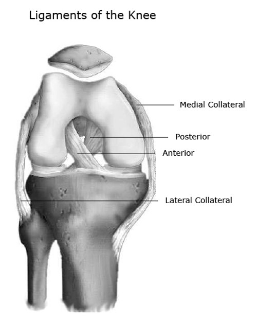
The four main ligaments in the knee connect the femur (thighbone) to the tibia (shin bone), and include the following:
Ligaments are flexible, but they do not stretch very far. Its complexity and its efficiency is the best example of god's creation. The severity of these symptoms depends on which ligament has been torn. You may be experiencing knee pain and want to know the possible causes. The knee is the largest joint in the body, and one of the most easily injured. They are associated with muscles discussed in the section above (see above). A diagram of the knee, including ligaments. Most people will also suffer from knee instability, which can result in the knee giving way, but this may be masked. The knee is designed to fulfill a number of functions: A complete tear will split the tendon into two pieces. The knee consists of three bones: It is held in place by a ligament at the bottom and a tendon on top. The largest joint in the body, the knee moves like a hinge, allowing you to sit, squat, walk or jump.
Bones, cartilage, ligaments, and tendons. Understanding the normal function of the knee joint can help you address some of these common. Knee pain could be the result of a problem with any one of these components, or a combination of several. Mcl & lcl found either side of the knee. Diagram of the ankle bones.
1 from Cross section of foot nerves 13 photos of the cross section of foot nerves cross section of nerve fiber, foot anatomy nerves, foot nerve pain, human foot nerves, nerve cross section histology, peripheral nerve cross section, spinal nerve cross section, foot, cross section of nerve fiber, foot anatomy. One of the most important tendons is the. There are two pairs of ligaments in the knee, collateral ligaments: Diagram of knee tendons and ligaments. Its complexity and its efficiency is the best example of god's creation. Acl & pcl found in the middle of the joint. The knee ligaments connect the thigh and shin bones (femur & tibia) and work together to control how the knee moves to keep it stable and prevent injury. The knee consists of three bones:
Around the knee there are two types of tendons.
Bones, cartilage, ligaments, and tendons. The knee joint is a complex structure that involves bones, tendons, ligaments, muscles, and other structures for normal function. The knee is the joint where the bones of the lower and upper legs meet. The tendons present in the knee are strong tissue bands that join the bones to the muscles. You may be experiencing knee pain and want to know the possible causes. The anterior cruciate ligament prevents the femur from sliding backward on the tibia (or the tibia sliding forward on the femur). Ligaments join the knee bones and provide stability to the knee: The knee consists of three bones: Diagram of inside the body. Diagram of knee tendons and ligaments. The muscles that affect the knee's movement run along the thigh and calf. The fdp laceration is usually treated with. Bones embedded in tendons are called sesamoid bones and they protect the tendons and improve the function of the joint by holding the tendons away from the center of the joint.
It connects the thigh bone to the shin bone. Tendons are elastic tissues made up of collagen. The anatomy of the knee consists of bones, muscles, nerves, cartilages, tendons and ligaments. Tendons are the connection between bones and muscles. The knee is the largest joint in the body, and one of the most easily injured.
Knee Joint Anatomy Bones Ligaments Muscles Tendons Function from www.healthpages.org The knee is the joint where the bones of the lower and upper legs meet. The knee ligaments connect the thigh and shin bones (femur & tibia) and work together to control how the knee moves to keep it stable and prevent injury. The largest tendon in the knee is the patellar tendon which covers the kneecap runs up the thigh and attaches to the quadriceps. It is made up of four main things: Mcl & lcl found either side of the knee. The four main ligaments in the knee connect the femur (thighbone) to the tibia (shin bone), and include the following: Understanding the normal function of the knee joint can help you address some of these common. The knee is the largest joint in the body, and one of the most easily injured.
Diagram of the ankle bones.
Ligaments are flexible, but they do not stretch very far. Three bones meet to form your knee joint: Related posts of knee tendon anatomy diagram and name chart cross section of foot nerves. Bones, cartilage, ligaments, and tendons. There are two pairs of ligaments in the knee, collateral ligaments: The medial or inside collateral ligament (mcl) connects the femur to the tibia. It is made up of four main things: Bones embedded in tendons are called sesamoid bones and they protect the tendons and improve the function of the joint by holding the tendons away from the center of the joint. Our interactive 3d knee diagram is an informative 360 degree rotating model. Jul 01, 2021 · free body diagram for calculating deltoid force. The ligament, located in the center of the knee, that controls rotation. Tendon diagram / knee tendons | skeletal | pinterest / a zone 1 injury involves an fdp tendon laceration distal to the fds insertion. A ligament is a type of fibrous tissue that usually.


0 Komentar